Images from a fetal MRI can provide information about conditions that might affect the mother or unborn baby. Images from a pelvic MRI can provide information about gynaecological conditions affecting women of all ages. Pelvic and fetal MRI have been widely used around the world for decades.
SO+GI Scan’s radiologists are qualified, specialist doctors with expertise in interpreting MRI images. We have more than 25 years of experience in ultrasound and MRI imaging specifically focused on fetal and pelvic images. We’ve supported many women and families through the MRI process and do our best to provide you with comfort, reassurance and timely answers.
Magnetic resonance imaging (MRI) is a non-invasive imaging technique that produces detailed 3D images of your body to detect health conditions and monitor treatment progress. It does not involve any radiation. It often creates clearer, more detailed images than an ultrasound.
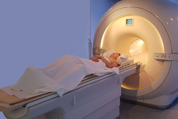
An MRI machine is essentially a large magnetic tube. It is housed in a specially designed, metal-free room and it creates images using magnetic fields and radio waves. Again, there is no radiation, just very large magnets.
MRI can be done on any part of the body and is particularly useful for soft tissues like your organs. At SO+GI Scan, we focus on MRIs to scan an unborn baby in the uterus (fetal MRI) and to identify or manage gynaecological conditions like fibroids and endometriosis (pelvic MRI).
The exact process of your MRI may vary depending on your personal situation and the reasons for having the scan. There may be some preparatory steps or the use of IV contrast (dye). We’ll inform you of exactly what to expect before you come to your appointment.
During an MRI, you lie inside a large magnetic cylinder, keeping very still to avoid blurring the image. The scan usually takes around 30-45 minutes.
To help you feel comfortable and to pass the time, we can:
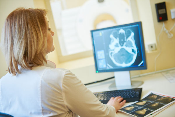
After the MRI, the images are reviewed by the specialist SO+GI scan radiologists.
A report will be sent to your doctors within the next few days.
Not everyone is comfortable in the closed-in space of an MRI machine.
If you would like more help to ease claustrophobia, please tell us. We may be able to give you some medicine that helps you relax.
Ultrasound is the standard way to assess your baby’s growth and development. If your ultrasound shows a suspected abnormality, we may recommend a fetal MRI. This can help us understand more about the abnormality and how it may affect the baby. Often, we will also recommend other tests or discussions with other specialists as part of your care.
An MRI of the fetus allows better views of some important anatomical structures than ultrasound alone. These areas include the fetal brain, spine, lungs and diaphragms, kidneys and bowel, and abdominal masses.
Fetal MRI can add more detail and clarity and give more information for genetic counselling, decision-making, and pregnancy management. It is particularly helpful in:
We understand how anxious you may feel about needing a fetal MRI. We’re here to support you throughout the process and to provide a timely report on the findings of your MRI scan.
MRI scans may be helpful additions to your ultrasound scans in gynaecology to clarify or confirm your underlying diagnosis. With its superior resolution and clarity, pelvic MRI may allow us to obtain clearer images of your pelvic organs and structures.
MRI also has a role in clarifying the anatomy and shape of a uterus and endometrial cavity in adolescents and sometimes in patients with infertility.
Some of the reasons a pelvic MRI can be used include to:
We understand how important gynaecological health is to your overall wellbeing and to your fertility. We treat you with sensitivity and care throughout your scan and ensure that we provide a timely report to your doctors.
Ultrasound is the standard way to assess your baby’s growth and development. If your ultrasound shows a suspected abnormality, we may recommend a fetal MRI. This can help us understand more about the abnormality and how it may affect the baby. Often, we will also recommend other tests or discussions with other specialists as part of your care.
An MRI of the fetus allows better views of some important anatomical structures than ultrasound alone. These areas include the fetal brain, spine, lungs and diaphragms, kidneys and bowel, and abdominal masses.
Fetal MRI can add more detail and clarity and give more information for genetic counselling, decision-making, and pregnancy management. It is particularly helpful in:

We understand how anxious you may feel about needing a fetal MRI.
We’re here to support you throughout the process and to provide a timely report on the findings of your MRI scan.

MRI scans may be helpful additions to your ultrasound scans in gynaecology to clarify or confirm your underlying diagnosis. With its superior resolution and clarity, pelvic MRI may allow us to obtain clearer images of your pelvic organs and structures.
MRI also has a role in clarifying the anatomy and shape of a uterus and endometrial cavity in adolescents and sometimes in patients with infertility.
Some of the reasons a pelvic MRI can be used include to:
We understand how important gynaecological health is to your overall wellbeing and to your fertility.
We treat you with sensitivity and care throughout your scan and ensure that we provide a timely report to your doctors.
CLICK HERE to learn more about MRIs.
If you have any further questions regarding fetal or pelvic MRI scans, please CONTACT US.