
Transabdominal Scan
Transvaginal Scan
Your doctor may refer you for a gynaecological ultrasound for one of these common indications:
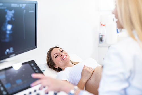
Vaginal ultrasound provides much clearer views and more detail of the pelvic structures. A narrow, gel covered probe is gently introduced into the vagina. The examination takes approximately 10-20 minutes.
In young girls, women who have not been sexually active or women who don’t feel comfortable having a vaginal ultrasound, a transabdominal ultrasound, or ultrasound through the abdomen can be performed. A very full bladder is necessary for the scan to be diagnostic.
Endometriosis is an often painful condition that can affect your fertility and quality of life.
Tissue that normally lines the inside of your uterus (the endometrium) grows outside of your uterus. This mainly happens on the wall of the pelvis, on the ligaments supporting the uterus and on the ovaries. Sometimes, it can also grow around your appendix, bowel, or bladder.
Like the endometrium inside the uterus, the endometrial implants swell and then bleed with your monthly hormonal cycle. Your body responds by surrounding the affected area with scar tissue (adhesions). This can result in damage to pelvic structures and may cause them to stick together. Over time, the endometrial tissue may also enlarge and form cysts, particularly in the ovaries.
Symptoms include pain during periods and with sexual intercourse. Some patients experience pain with ovulation, urination or defaecation with periods. Other patients present with abnormal bleeding or infertility.
While superficial lesions of endometriosis cannot be reliably diagnosed with ultrasound, deep infiltrating endometriosis usually causes more distortion of normal anatomy in your pelvis. It can infiltrate into ligaments, bowel and bladder forming nodules and adhesions which can often be detected with transvaginal ultrasound.
A comprehensive transvaginal scan is performed, and results will be discussed with you either during or immediately after the scan by one of our specialists, and a detailed report will be provided to your doctor.
A magnetic resonance imaging (MRI) scan can detect endometriosis sites deep within your pelvis, which can be difficult to see with ultrasound alone. Identifying and mapping these sites enables your doctors to develop a surgical plan to remove them and relieve your symptoms.
Saline Infusion Sonography (SIS) is an ultrasound-guided technique designed to better image the uterine cavity. A thin catheter is introduced carefully through the cervix and a small amount of sterile saline is injected through the catheter to allow for more accurate assessment of the uterine cavity.
HyCoSy is a transvaginal ultrasound technique used in the investigation of infertility. Its primary aim is to establish whether or not the woman’s fallopian tubes are patent or whether one or both is blocked.
The technique involves introducing an ultrasound contrast agent (Ex Em Foam) injected into the uterine cavity, which is then observed as it flows through the fallopian tubes to assess tubal patency. The contrast generates bright echoes that make visualisation of the tubes easier.
HyCoSy has increasingly been used as a first line investigation for infertility because it is convenient and safe.
A sterile speculum is placed in the vagina, and a thin catheter is then inserted into the uterine cavity through the cervical canal. The speculum is then replaced by a transvaginal ultrasound probe. The bright echoes generated by the contrast enable us to visualise the fallopian tubes and determine if there are any blockages along the way, the image is further improved by the addition of colour Doppler imaging. We use a normal sterile saline first to check for any issues (SIS), before using the ExEm foam contrast.
The test is performed from Day 7 to 10 of your typical menstrual cycle, to make the uterine lining easier to assess and to ensure there is no risk of a potential pregnancy.
It does not refer to bleeding in pregnancy, which is likely to have a different cause and need a different response.
There are many potential causes of abnormal uterine bleeding, including:
Bleeding or spotting that occurs after you have stopped having menstrual periods.
Common causes include inflammation and thinning of the lining of the vagina (atrophic vaginitis) and growths in the uterus or cervix (polyps) which are usually benign.
The bleeding may be due to endometrial cancer in a very small number of cases which is why it is important not to ignore it. Transvaginal ultrasound is a very effective way to look for any polyps or areas of thickening in the uterine lining (endometrium).